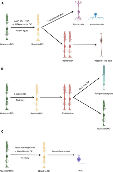
Schematic illustration of MG-derived in vivo neurogenesis in the adult mouse retina. (A) MG-derived regeneration of bipolar or amacrine-like cells. In the presence of NMDA-induced injury, Ascl1 overexpression combined with TSA or Nfi deletion combined with growth factors reprograms MG into two clusters of cells. One cell cluster is composed of bipolar and amacrine-like cells, and the other cell cluster contains progenitor-like cells that are mostly non-neurogenic. (B) MG-derived regeneration of rod photoreceptors. In the absence of retinal injury, β-catenin overexpression stimulates MG to reenter the cell cycle to proliferate as the first step. A subsequent gene transfer of Otx2/Crx/Nrl induces one daughter cell to differentiate into a rod photoreceptor, while the other daughter cell remains a quiescent MG. (C) MG-derived regeneration of RGCs. In the absence of retinal injury, Ptbp1 downregulation or Math5/Brn3b overexpression converts MG into RGCs by direct transdifferentiation. However, genetic-based fate mapping experiments, independent of AAV-mediated gene transfer, were not performed in these studies to trace the lineage of MG after Ptbp1 downregulation or Math5/Brn3b overexpression. OE, overexpression; GF, growth factors. Created with BioRender.com.
|
