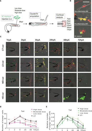
(A) Experimental design of macrophage recruitment, and activation following infection and amputation. (B) Legends (Z projections of confocal images) to distinguish non-inflammatory macrophages (red) in Tg(mpeg1:mCherry-F) larvae from pro-inflammatory (orange) macrophages in (Tg(mpeg1:mCherry-F;tnfa:eGFP-F) larvae (Scale bar = 30 µm). Wasabi-expressing M. marinum are in green. (C) Z projections of confocal images allowed to establish the kinetic of recruitment and activation of macrophages after injection of PBS (CT) or infection with LD, MD or HD of M. marinum at 1, 3, 6, 24, 48, 72 hpA (Scale bar = 100 µm). (D) Kinetic of total macrophages recruited after fin amputation following injection of PBS (CT) or infection with LD, MD or HD of M. marinum (mean ± SEM, n< 30, Kruskal-Wallis, Dunn’s multiple comparisons test, *p ≤ 0.05, **p ≤ 0.01, ****p ≤ 0.0001). (E) Kinetic of pro-inflammatory macrophages recruitment (expressed as the fold change of mpeg+ and tnfa+ macrophages over the total number of mpeg+ macrophages) after fin amputation following injection of PBS (CT) or infection with LD, MD or HD of M. marinum (mean ± SEM, n<30, Kruskal-Wallis, Dunn’s multiple comparisons test, *p ≤ 0.05, ****p ≤ 0.0001).
|
