
Figure 7
- ID
- ZDB-FIG-220409-53
- Publication
- Gupta et al., 2022 - Cilia proteins are biomarkers of altered flow in the vasculature
- Other Figures
- All Figure Page
- Back to All Figure Page
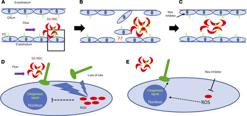
Proposed mechanism of loss of cilia proteins. The schematic demonstrates cilia stability mechanisms associated with sickle RBCs (SS RBCs) and brain ECs in an SCD setting. Green-colored structures on RBCs and ECs are cilia, and red-colored dots in (D and E) are oxidative stress components, such as ROS. SS RBCs create altered shear stress on ECs due to their sickle shape. Upon interaction with ECs, SS RBCs elevate endothelial oxidative stress (A and D). The physical (shear) or metabolic (oxidative) stress results in loss of endothelial cilia or cilia-associated proteins. These shredded cilia fragments (and associated proteins) are now available for interaction with SS RBCs and may enrich the RBCs’ surface (B). Upon attenuation of oxidative stress in ECs (NOX inhibitor), the loss of endothelial cilia or any subsequent enrichment of cilia in sickle RBCs is minimized (C, fewer green cilia in RBC compared with in B). (D and E) Magnified images of A, which details the mechanism identified in this study. Sickle RBCs upon adhering to ECs elevate oxidative stress (increased ROS) in ECs (D). This causes loss of cilia or cilia-associated protein and may send inhibitory signals to the nucleus to prevent a ciliogenesis signal. Once the oxidative stress is attenuated by NOX inhibition, ROS is downregulated, and restoration of endothelial cilia is observed (E). |