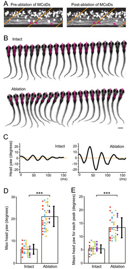
Ablation of MCoD neurons lead to the increase of head-yaw displacement during spontaneous swimming. (A) Confocal stacked images of Tg[evx2-hs:GFP] fish before (left) and after (right) laser ablation. Images of two hemi-segments are shown. Magenta arrows show MCoD neurons that were chosen for laser ablation. MCoD neurons can be identified by their very ventral location in the spinal cord. Brown lines show boundaries of muscle segments. Scale bar, 20 μm. (B) Successive images captured at 1000 frames per second of larval zebrafish swimming. Images of every three frames (3 ms interval) are shown. Magenta bars depict the head directions in each frame. Top, images of an intact fish. Bottom, images of an MCoD-ablated fish. Scale bar, 500 μm. (C) Graphs of head yaw angle (y axis) versus time (x-axis) during swimming. Left, intact fish. Right, MCoD-ablated fish. (D) Maximum head yaw angle of intact and MCoD-ablated fish during swim bouts. Five fish were examined for each fish type. For each fish, 10 swim bouts were examined. Data obtained from the same fish are color coded. (E) Mean head yaw angle for displacement peaks of intact and MCoD-ablated fish during swim bouts. Five fish were examined for each fish type. For each fish, 10 bouts were examined. Data obtained from the same fish are color coded (the same fish as D). ***p < 0.001 (two-tailed t-test).
|

