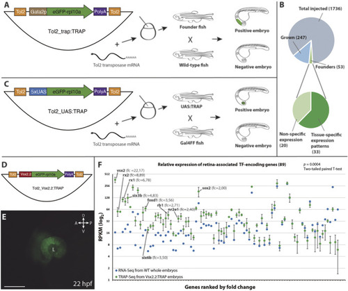
Vectors and procedures used to expand TRAP approaches in zebrafish.(A): The Tol2_trap:TRAP vector comprises a cassette containing the eGFP-rpl10a fusion gene (green) and the gata2p minimal promoter (brown), flanked by Tol2 recognition sequences (orange). This vector was injected together with Tol2 transposase mRNA in one-cell stage zebrafish embryos. Once grown, adult fish were screened for eGFP-rpl10a expression in their progeny. (B): Pie charts showing the efficiency rate of the trap:TRAP approach. (C): The Tol2_UAS:TRAP vector comprises a cassette that contains the eGFP-rpl10a fusion gene (green) together with the 5xUAS element (blue), flanked by the Tol2 recognition sequences (orange). This vector together with Tol2 transposase mRNA were injected together in one-cell stage embryos. Once grown, adult fish were outcrossed with a Gal4 line to identify founders. (D): Schematic representation of the Tol2_vsx2.2:TRAP vector used to test the functionality of the eGFP-rpl10a cassette (green) under the control of the vsx2.2 promoter (red). (E): Retina-specific eGFP-rpl10a expression in the line Tg[vsx2.2:TRAP] tested in TRAP-seq experiments at 22 hpf. L, lens. Scale bar = 100 µm. (F): Differential expression (RPKM) of retinal TF-encoding genes in affinity-purified transcripts from vsx2.2:TRAP embryos at 22 hpf (TRAP-seq, n = 3, green), and in 22 hpf whole embryos RNA-seq sample (control, blue). Note the significant expression enrichment of neural retina-associated transcription factors (ranked by fold-change) in the vsx2.2:TRAP sample (p = 0.0004; two-tailed paired t-test). RPKM values corresponding to TFs from the core retinal GRN, as well as their fold change vs the control, are indicated.
|
