
Fig. 5
- ID
- ZDB-FIG-220204-20
- Publication
- Wong et al., 2021 - Loss of C2orf69 defines a fatal autoinflammatory syndrome in humans and zebrafish that evokes a glycogen storage-associated mitochondriopathy
- Other Figures
- All Figure Page
- Back to All Figure Page
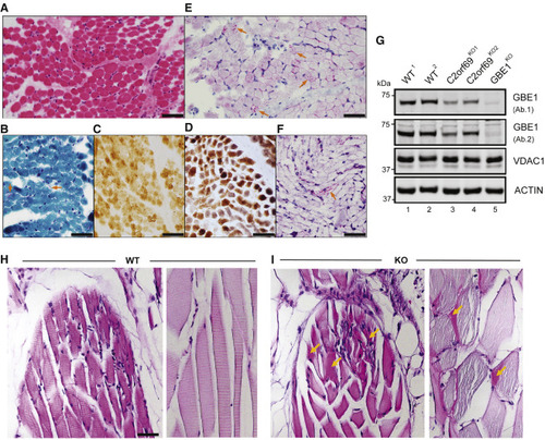
(A) Skeletal muscle biopsy of proband II:3 from family 8 shows mild variation of fiber size in H&E sections. (B) Subsarcolemmal accumulation of mitochondria is apparent in some fibers (arrows) by modified Gomori-trichrome stain. (C) Several fibers show faint staining by cytochrome-c-oxidase (COX) stain. (D) These COX-deficient fibers are more easily identified as bluish fibers by COX-SDH stain. (E) Periodic acid Schiff (PAS) stain shows unusual granular staining in many fibers (arrows), which are partially resistant to diastase, as seen by PAS-diastase stain (F). (F) PAS-positive material is seen in a few fibers after diastase treatment. Scale bars represent 50 μm. (G) Approximately 50% decrease in endogenous GBE1 levels is seen in two independent C2orf69 KO HAP1 cell lines with two different antibodies (Ab1, Proteintech; Ab2, Abcam). Two WT and GBE1 KO HAP1 cell lines act as positive and negative controls. Antibodies against VDAC1 and ACTIN are used as loading controls. (H) PAS stain in skeletal muscles of WT zebrafish at 6 months of age. (I) PAS-positive aggregates (marked by orange arrows) in skeletal muscles of C2orf69 KO zebrafish are suggestive of glycogen accumulation. Sarcomeres were partially disorganized in mutant striated muscle fibers. Scale bar represents 50 μm. |