
Figure 2
- ID
- ZDB-FIG-220104-168
- Publication
- Piccione et al., 2021 - STW 5 Herbal Preparation Modulates Wnt3a and Claudin 1 Gene Expression in Zebrafish IBS-like Model
- Other Figures
- All Figure Page
- Back to All Figure Page
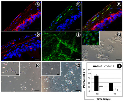
Immunofluorescent staining showing the expression of (A) Frizzled 9 (red) within the epithelial and neuromuscular compartments; (B) PAN neuronal marker (green) within the neuromuscular layer; and (C) both markers (Frizzled 9 and PAN neuronal marker) in colocalization (yellow); and (D) Wnt3a within the epithelium and neuromuscular layer. Nuclear counterstaining was performed using DAPI (blue). (E) Whole-mount immunofluorescent staining of zebrafish ENS network by detection of PAN neuronal marker (green) Bar: 50 µm. Optical microscopy of (F) primary intestinal epithelial cells, characterized by the expression of Pan-cytokeratin (left corner, green); ENS cultures at (G) 24 h and (H) 7 days from isolation. Bar: 100 µm (200 µm in left corner). (I) Flow cytometry analysis of Sox2 and Sox10 in ENS cells at the time of isolation (T0) and after culturing for 7 days (T7). Data are expressed as percentage (%) of positive cells ± standard deviation (SD). Created with BioRender.com, accessed on 31 October 2021. |