
Figure 1 -
- ID
- ZDB-FIG-211210-5
- Publication
- Ravenscroft et al., 2021 - Heterozygous loss-of-function variants significantly expand the phenotypes associated with loss of GDF11
- Other Figures
- All Figure Page
- Back to All Figure Page
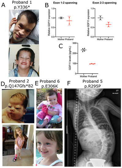
Fig. 1. Overview of patients with GDF11 variants. (a) Pictures of proband 1. (b) GDF11 expression was measured in peripheral blood mononuclear cells (PBMCs) derived from the proband or unaffected mother by quantitative polymerase chain reaction (qPCR) using primer sets spanning exons 1 and 2 (left) or 2 and 3 (right) normalized to GUSB loading control expression. RNA was collected from n = 2 technical replicates from N = 1 blood draws per patient. Error bars = SD. (c) GDF11 expression was measured in plasma derived from the proband or unaffected mother using a commercial GDF11 enzyme-linked immunosorbent assay (ELISA) kit (LSBio #LS-F11519) Error bars = SEM. Quantification was performed in n = 4 technical replicates from N = 1 blood draw per patient. Pictures of proband 2 (d) and proband 6 (e). X-ray of proband 5 (f). |