
Fig. 5
- ID
- ZDB-FIG-211118-98
- Publication
- Abu Nahia et al., 2021 - Genomic and physiological analyses of the zebrafish atrioventricular canal reveal molecular building blocks of the secondary pacemaker region
- Other Figures
- All Figure Page
- Back to All Figure Page
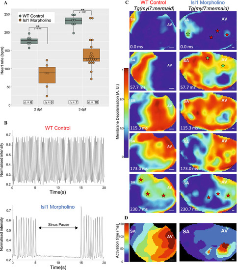
Effects of morpholino knockdown of isl1 on electrical activity of the atria in zebrafish larvae. A Heart rate at 48 hpf (left) and 72 hpf (right) in WT control (grey) and isl1 morphant (brown) zebrafish, showing slowed heart rate in isl1 morphants (Wilcoxon rank sum test with continuity correction, asterisks indicate p value ≤ 0.01). B Videographic analysis of the heartbeat in WT control (top) and isl1 morphant (bottom) 48 hpf zebrafish, showing slowed heart rate and sinus pauses (during the period indicated by the arrow) in the isl1 morphant. C Sequence of video frames showing electrical activation of the atria in WT control (left) and isl1 morphant (right) 48 hpf Tg(myl7:mermaid) zebrafish, showing normal activation (from the sinoatrial node region [SA] to the atrioventricular [AV] junction) and sites of latest activation/repolarisation (indicated by red stars) in the WT control and abnormal activation in the isl1 morphant (sites of early/ectopic activation in the morphant zebrafish indicated by green stars). D Isochronal activation map of the atria in WT control (left) and isl1 morphant (right) 48 hpf zebrafish derived from the video represented in (C) |