
Figure 1.
- ID
- ZDB-FIG-211111-37
- Publication
- Jin et al., 2021 - egfl6 expression in the pharyngeal pouch is dispensable for craniofacial development
- Other Figures
- All Figure Page
- Back to All Figure Page
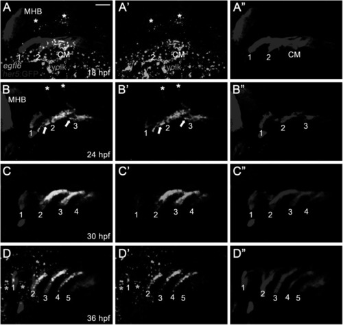
Expression of egfl6 in pouch formation. (A-D) Fluorescence in situ hybridization of egfl6 (green) in conjunction with the GFP immunohistochemistry (red) in wild-type Tg(her5:GFP) animals. (A) At 18 hpf, egfl6 expression is seen in the her5-positive second (2) pouch and the posterior cell mass (CM), with no egfl6 expression seen in the first (1) pouch. egfl6 expression is also seen in the developing hindbrain (asterisks). Note of non-specific green staining in the yolks (dotted line). (B) At 24 hpf, egfl6 is expressed in all three her5-positive pouches (1-3), with new egfl6 expression appearing in the mesoderm between pouches (arrows). egfl6 expression is still seen in the developing hindbrain (asterisks). (C) At 30 hpf, egfl6 expression is only observed in all four her5-positive pouches (1-4), with the egfl6 expression in the mesoderm gone. (D) At 36 hpf, egfl6 is expressed in all pouches, with its expression in the fifth (5) pouch being faint. Also, unidentified tissues adjacent to the first (1) and second (2) pouches express egfl6 (asterisks). Note that the sixth pouch is barely seen at the level of tissues. MHB: midbrain−hindbrain boundary. (A′–D′) Green channel only. (A”–D”) Red channel only. Scale bar: 40 μm. |
| Gene: | |
|---|---|
| Fish: | |
| Anatomical Terms: | |
| Stage Range: | 14-19 somites to Prim-25 |