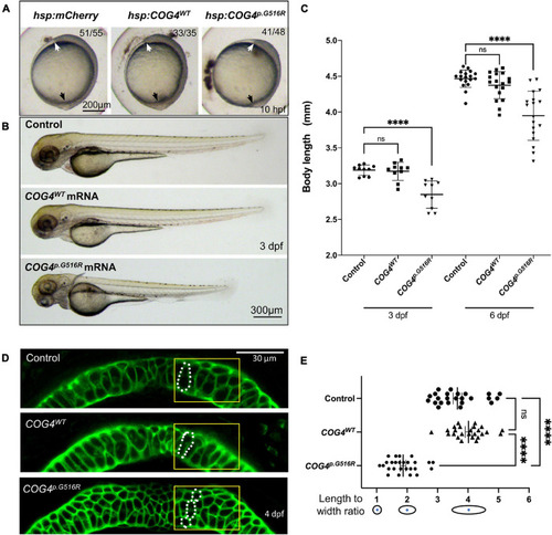
Expression of human COG4p.G516R impairs zebrafish early development and chondrocyte intercalation. (A,B) Expression of COG4p.G516R in zebrafish causes gastrulation defects and shortened body axis. (A) Lateral view of representative embryos. Anterior to the top. Compared to the control construct hsp70l:mCherry and COG4WT, embryos expressing COG4p.G516R show an axis extension defect at 10 hpf. White arrow points to the head region, and the black arrow points to the tailbud. Expression of COG4p.G516R mRNA causes similar results. (C) Graphs show the measured body length of each group at 3 and 6 dpf. The data are presented as mean ± SD. One-way ANOVA with Tukey’s multiple comparison tests was applied. ****p < 0.0001; ns, not significant. (D) Expression of COG4p.G516G causes craniofacial abnormalities. Ventral view of representative Meckel’s cartilage of zebrafish larvae at 4 dpf after WGA staining and imaged by a confocal microscope. Dotted circular lines highlight chondrocyte cell shape and their relative configuration with each other. (E) Graphical representation of the length-to-width ratio of chondrocytes in the region of interest, yellow box in (D). Individual cell length-to-width ratio was measured in three representative Meckel’s cartilage images of each group. The data are presented as mean ± SEM. One-way ANOVA with Tukey’s multiple comparison tests was applied. ****p < 0.0001; ns, not significant. Experiments were performed in triplicates with similar results.
|
