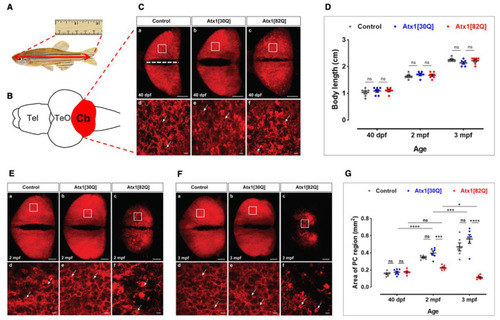
Progredient disintegration of PC layer in genetic model of SCA1 in zebrafish. (A) Schematic drawing of adult zebrafish to demonstrate measurement of body length form the anterior tip of the jaw until the caudal indentation of the tail fin. (B) The cerebellum in dissected brains from these specimens is easy to identify by the red fluorescent PC population forming a continuous layer of cells across the corpus cerebelli (dorsal view). (C) Maximum brightness projection of images stacks recorded by confocal microscopy from control (a,d), Atx1[30Q] (b,e), and Atx1[82Q] (c,f) expressing heterozygous carriers of the respective transgene at 40 dpf. Note speckles of brighter fluorescence suggestive of condensation and cellular shrinkage can be observed in the PC layer of Atx1[82Q] carriers not found in GAPmScarlet and Atx1[30Q] controls (compare white arrows). (D) Body length measurements reveal a similar overall growth rate to control (n = 6), Atx1[30Q] (n = 6), and Atx1[82Q] (n = 6) fish. (E) Maximum brightness projection of image stacks recorded by confocal microscopy from control (a,d), Atx1[30Q] (b,e), and Atx1[82Q] (c,f) expressing heterozygous carriers of the respective transgene at two months of age and (F) three months of age, respectively. The area marked by a white rectangle is displayed at higher magnification below for each specimen, respectively. Compared to controls and Atx1[30Q], young adult Atx1[82Q] zebrafish at three months of age display a clear atrophy of their PC layer containing widespread fluorescent debris of degenerated PCs (Ff white arrows). (G) Quantification of area covered by fluorescent PCs, while in controls (n = 6) and Atx1[30Q] carriers (n = 6) a continuous age-dependent increase of the PC layer occurs, the expansion of the PC layer in Atx1[82Q] carriers (n = 6) stalls. (C,E,F): dorsal views of corpus cerebelli, anterior to the left, scale bars: 100 µm (a–c) or 10 µm (d–f). The data in (D,G) are presented as mean ± SEM, * p < 0.05, *** p < 0.001 and **** p < 0.0001 according to two-way ANOVA with post hoc Tukey’s test. n = 7, 6, 6 in 40 dpf, 2 mpf (months postfertilization) and 3 mpf control groups, respectively. n = 7, 6, 6 in 40 dpf, 2 mpf, and 3 mpf Atx1[30Q] groups; and n = 6, 7, 7 in 40 dpf, 2 mpf, and 3 mpf Atx1[82Q] groups, respectively. Abbr.: Cb: cerebellum, PC: Purkinje cell, TeO: optic tectum, Tel: telencephalon.
|
