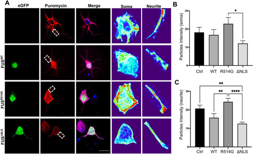
Overexpression of FUS mutations alters protein translation within transfected neurons. (A) Representative images of primary cortical neurons transfected with eGFP-FUSWT, eGFP-FUSR514G or eGFP-FUSΔNLS (green) and stained for puromycin (red) and MAP2 (merge). Zoomed in images of each soma and neurite which were analysed has been included for each condition and presented in a heatmap to show changes more clearly. (B) Quantification of average intensity of puromycin puncta within soma show a non-significant decrease when eGFP-FUSΔNLS is compared to eGFP-FUSWT (p = 0.5704). Comparison of eGFP-FUSWT to control showed no significance (p = 0.2041) whereas comparison of eGFP-FUSWT to eGFP-FUSR514G showed a non-significant increase (p = 0.4042). However, comparison of eGFP-FUSR514G to eGFP-FUSΔNLS showed significant decrease (p < 0.05). (C) Quantification of protein translation in neurites showed a significant increase when eGFP-FUSΔNLS or eGFP-FUSwt was compared to eGFP-FUSR514G (P < 0.0001 and p < 0.01 respectively). Furthermore, a significant decrease was observed when control was compared to both eGFP-FUSΔNLS (P < 0.01) while a non-significant decrease was seen for comparison against and eGFP-FUSwt (p = 0.2041). When eGFP-FUSΔNLS was compared to eGFP-FUSwt, no significant change was observed (P = 0.5704). Statistical analysis was performed using a One-Way ANOVA with a post-hoc Tukey’s multiple comparisons test; error bars are ± SEM. N = five cells were analysed from three different independent replicates. *p < 0.05, **p < 0.01, ***p < 0.001 ****p < 0.0001.
|
