
Figure 2
- ID
- ZDB-FIG-210707-120
- Publication
- Alexandre-Moreno et al., 2021 - Null cyp1b1 Activity in Zebrafish Leads to Variable Craniofacial Defects Associated with Altered Expression of Extracellular Matrix and Lipid Metabolism Genes
- Other Figures
- All Figure Page
- Back to All Figure Page
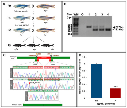
Generation and molecular characterisation of a cyp1b1-KO zebrafish line using CRISPR/Cas9 genome editing. (A) Stepwise procedure followed to generate the KO line. Adult F0 zebrafish were crossed with wildtype AB animals and the offspring were genotyped by PCR and agarose gel electrophoresis to identify germline transmission of cyp1b1 deletions (F0 founders). The selected F0 founder was mated with a wildtype AB animal to obtain mutant F1 heterozygotes that were further outcrossed to segregate off-target mutations in the F2 generation. F2 heterozygotes were inbred to obtain F3 fishes. The scheme was created with the Biorender tool (https://biorender.com/) (accessed on 21 April 2021). (B). Genotyping by PCR and agarose gel electrophoresis of a 133-bp cyp1b1 deletion in the F3 generation. Representative examples of the three genotypes are shown. Black arrowhead: wildtype allele (372 bp). White arrowhead: mutant allele (239 bp). (C) Sanger sequencing of the selected mutation. The top diagram indicates localization of the identified deletion in cyp1b1 exon 1. Scissors: DNA cleavage sites targeted by the two crRNAs. The numbers correspond to cDNA nucleotide positions. Red arrows in the electropherograms indicate the 5′ and 3′ ends of the deletion. Deleted nucleotides are indicated in the red background. The diagram in the bottom represents the mutant exon. (D) Decreased mRNA levels in cyp1b1-KO zebrafish. Pools of 45 F4 zebrafish larvae (48 hpf) were used to quantify relative cyp1b1 mRNA levels by RT-qPCR. The values represent the average of three independent experiments carried out in triplicate. Asterisks indicate statistical significance compared to the wild type, p < 0.001 (***). |