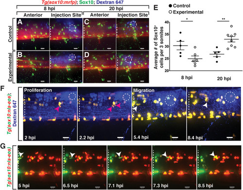
Lysolecithin alters the number and behavior of Sox10+ cells in the spinal cord. All images are lateral views of the spinal cord with anterior to the left and dorsal to the top. (A–D) 4 dpf Tg(sox10:mrfp) zebrafish larvae injected with either a control solution [control, (A,C)] or lysolecithin [experimental, (B,D)] containing the fluorescent tracer Dextran-647 to identify the injection site. Injected zebrafish were fixed at 8 hpi (A,B) or 20 hpi (C,D) and labeled with an antibody to Sox10 (green). (E) Quantification of the average number of Sox10+ cells within the injection site and spanning the width of approximately three motor nerves to capture the effects from lysolecithin (experimental) or control solutions. *p = 0.0252 at 8 hpi (n = 5 control; n = 6 experimental) and **p = 0.0033 at 20 hpi (n = 5 control; n = 8 experimental). Statistical significance was measured using an unpaired t-test. (F) Time-lapse imaging of proliferation (red arrowhead) and migration (white arrowhead) of sox10+ cells in a Tg(sox10:nls-eos) zebrafish larva following injection of lysolecithin at 4 dpf. White dashed ellipse denotes the injection site. (G) Time-lapse movie following injection of lysolecithin and photoconversion of the lesion area in a 4 dpf Tg(sox10:nls-eos) zebrafish larva. Red fluorescence denotes the photoconverted cells within the lesion. Green fluorescence denotes the cells outside of the lesion. Arrowhead identifies a sox10+ cell anterior to the injection site that migrates posteriorly into the drug dispersal region. White dashed lines denote the spinal cord. Scale bars, 25 μm.
|
