
Fig. 6
- ID
- ZDB-FIG-210520-63
- Publication
- Wang et al., 2021 - Eukaryotic initiation factor 4A3 inhibits Wnt/β-catenin signaling and regulates axis formation in zebrafish embryos
- Other Figures
- All Figure Page
- Back to All Figure Page
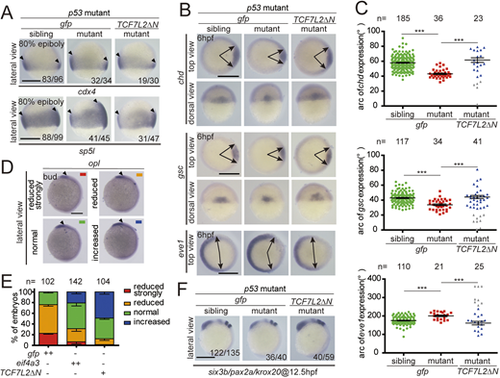
(A) Expression areas of Wnt targets in indicated groups of embryos at 80% epiboly stage. (B) Expression of the dorsoventral markers in indicated groups at 6 hpf. (C) Quantification of the arc of marker expression shown in B. A small number of mutant embryos within the TCF7L2ΔN mRNA injected groups showed more severe phenotypes in comparison with those of mutant embryos injected with gfp mRNA (gray data points). (D,E) Eif4a3 and TCF7L2ΔN rescue forebrain defects in apc mutants. Representative expression domain of the anterior neural marker, opl, in each indicated class of embryos at the bud stage (D) and quantification of each indicated class (E). One-cell stage embryos of apc+/−×apc+/− were injected with 200 pg of gfp or eif4a3 mRNA, or 50 pg of TCF7L2ΔN mRNA, raised to the bud stage and subjected to whole-mount in situ hybridization analysis. Each embryo was photographed and then genotyped. (F) Expression of anteroposterior neural markers in each indicated group of embryos at 12.5 hpf. One-cell stage embryos were injected with 50 pg of gfp or TCF7L2ΔN mRNA, raised to the indicated stage and subjected to whole-mount in situ hybridization analysis. Each embryo was photographed and then genotyped. All embryos are lateral views with dorsal to the right and anterior upwards, top views with dorsal to the right, and dorsal views with animal pole upwards. Embryos are examples from three pairs of adult fishes. Data are mean±s.e.m. ***P<0.001. Unpaired t-test, two-tailed. Scale bars: 200 μm. |