
Fig. 4
- ID
- ZDB-FIG-210513-4
- Publication
- Banavar et al., 2021 - Mechanical control of tissue shape and morphogenetic flows during vertebrate body axis elongation
- Other Figures
- All Figure Page
- Back to All Figure Page
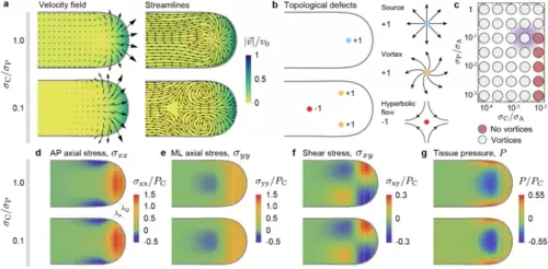
Morphogenetic flows and stress field during unidirectional elongation of the body axis. (a,b), Velocity field and streamlines of morphogenetic flows for different values of 𝜎C/𝑃𝐶, showing two structurally different flow fields (a). For large capillary stresses 𝜎C, the flow emerges from a single topological defect (a; source) of charge +1 (b). For lower capillary stresses, the tissue flow dramatically changes its structure and is characterized by three topological defects, namely 2 counter-rotating vortices (each with charge +1) and a stagnation point associated with hyperbolic flow (with charge −1). The remaining parameter values for each case are those indicated in Fig. 2a for unidirectional elongation (UE) in the absence and presence of vortices, labelled nv (no vortices) and v (vortices), respectively. (c), Diagram indicating the structure (presence or absence of vortices) of tissue flows. The tissue morphogenetic flows sharply transit from the two flow structures shown in (a) as the parameters are changed. The diffuse violet color indicates the estimated parameter region for wild type zebrafish body axis elongation. (d–g), Spatial distribution of all components of the stress tensor, namely the AP axial stress 𝜎𝑥𝑥 (d), the mediolateral stress 𝜎𝑦𝑦 (e) and the shear stress 𝜎𝑥𝑦 (f), as well as the tissue pressure P (g), for the two examples of tissue flows shown in panel (a). Despite the dramatically different structures of the flow field, the stresses are very similar. Both the location of the fluid-to-solid transition and the size of the region where cells enter ventral tissues are indicated by gray dashed lines at distances 𝜆𝜇 and 𝜆𝑄, respectively, from the posterior body end. |