FIGURE

Fig. 4

ID
ZDB-FIG-210512-39
Publication
Tan et al., 2021 - Small Extracellular Vesicles Deliver TGF-β1 and Promote Adriamycin Resistance in Breast Cancer Cells
Other Figures
All Figure Page
Back to All Figure Page
Fig. 4

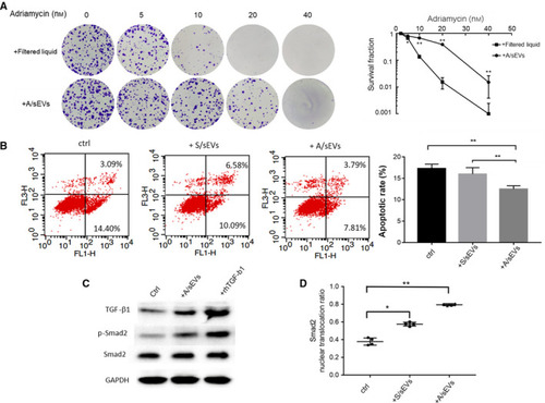
The effects of sEVs on inhibition of apoptosis in the recipient cells. (A) The isolated A/sEVs were finally purified using a specific column to remove soluble molecules accompanied by sEVs, and the eluted sEVs and filtered liquids were incubated with MCF‐7 cells. After treated with adriamycin as indicated, the cell survival rate was determined by clonogenic assay. (B) MCF‐7 cells were incubated with S/sEVs and A/sEVs and then treated with adriamycin, and the apoptotic cells were quantified by flow cytometry. The reduction in apoptosis by uptake of sEVs was calculated. **P < 0.01 represents the significances between the two groups as indicated. (C) The levels of TGF‐β1 and relating phosphorylated Smad2 in the recipient cells were quantified by western blots. Treatment with rhTGF‐β1 protein was used as a positive control. (D) Nuclear and cytoplasmic Smad2 proteins in the sEV uptake cells were measured using a Smad2 ELISA Kit. The percentage of nuclear‐translocated Smad2 was calculated by normalized with total Smad2. All experiments were repeated, and more than 3 replicates were included in A, B, and D. The data were presented as the mean ± standard deviation. *P < 0.05 and **P < 0.01 represent the significances between two groups as indicated.

Expression Data

Expression Detail
Antibody Labeling
Phenotype Data

Phenotype Detail
Acknowledgments
This image is the copyrighted work of the attributed author or publisher, and ZFIN has permission only to display this image to its users. Additional permissions should be obtained from the applicable author or publisher of the image. Full text @ Mol. Oncol.