
Figure 1
- ID
- ZDB-FIG-210503-170
- Publication
- Suárez et al., 2021 - Cells with Many Talents: Lymphatic Endothelial Cells in the Brain Meninges
- Other Figures
- All Figure Page
- Back to All Figure Page
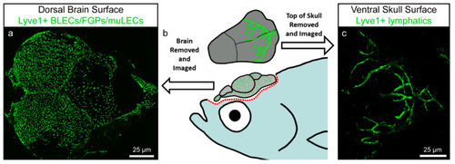
Zebrafish brain lymphatic endothelial cells (BLECs)/Fluorescent Granular Perithelial cells (FGPs)/mural lymphatic endothelial cells (muLECs) and meningeal lymphatic vessels in adult specimens. Image modified from Castranova et al. [49]. (a) Dorsal confocal image showing BLECs/FGPs/muLECs extended over the whole brain surface. (b) Schematic representation of an adult zebrafish depicting the dissection procedure for imaging of BLECs/FGPs/muLECs and intracranial meningeal lymphatic vessels. (c) Confocal image of the inner surface of the skull showing the meningeal lymphatic vessels that stay attached to the skull after dissection. |