
Fig. 4
- ID
- ZDB-FIG-210419-32
- Publication
- Cai et al., 2021 - Identification and functional characterization of the transcription factor coding Dp1 gene in large yellow croaker Pseudosciaena crocea
- Other Figures
- All Figure Page
- Back to All Figure Page
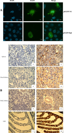
Subcellular localization of PcDp1. (A) Subcellular colocalization of PcDp1 protein analyzed by a GFP reporter assay. COS-7 cells were transfected with GFP reporter constructs expressing an EGFP-tagged Dp1. Empty pEGFP-N1 vector was used as a control. Nuclear DNA was stained with DAPI and cells were analyzed with a fluorescence confocal microscope. Scale bars, 10 μm. (B) Immunohistochemical localization (chocolate brown) of PcDp1 in tissues of health large yellow croaker. The specimens were fixed in 10% buffered formalin, paraffin embedded and sectioned. After antigen retrieval, sections were stained with rabbit polyclonal PcDp1 antisera or control sera, followed by HRP-coupled secondary Ab and visualized by the DAB method. Sections were visualized at 10×100 magnification. The specificity of the PcDp1 staining was confirmed by negative staining patterns observed with the normal rabbit serum staining. Note: 1,2-sections of spleen; 3,4-sections of head kidney; 5,6-sections of kidney; 7,8-sections of gills; 1,3,5,7-control sections; 2,4,6,8-positive sections. Arrows indicate areas of positive reaction in cytoplasm. |