
Fig. 5
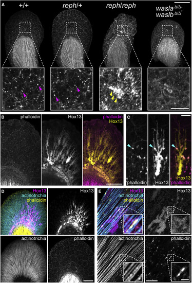
Figure 5. F-actin formation in the early fin fold is affected in wasl mutants and is associated with Hox13-positive cells and the actinotrichia (A) 3 dpf phalloidin-labeled pectoral fins showing modified F-actin in reph homozygous mutants and wasla; waslb double mutants. reph homozygotes develop large F-actin aggregates and dysmorphic fin folds, and wasla; waslb double mutants display a more dispersed F-actin network. (B) F-actin foci colocalize with the extensions of Hox13-positive cells in the fin fold marked with the Tg(2PΔepi:eGFP) reporter transgene. (C) A single Tg(2PΔepi:eGFP) Hox13-positive cell showing F-actin colocalization. (D) Triple labeling demonstrates colocalization of F-actin foci, Hox13-positive cells expressing the Tg(mInta-11:mCherry) transgene, and actinotrichia in the fin fold. (E) A single Tg(mInta-11:mCherry) Hox13-positive cell showing colocalization with actinotrichia fibrils and F-actin. Anterior to left, distal to top in all panels; scale bars (A) 100 μm (25 μm inset), (B, D) 50 μm, (C, E) 10 μm (2 μm inset in E). |
| Fish: | |
|---|---|
| Observed In: | |
| Stage: | Protruding-mouth |
Reprinted from Cell, 184(4), Hawkins, M.B., Henke, K., Harris, M.P., Latent developmental potential to form limb-like skeletal structures in zebrafish, 899-911.e13, Copyright (2021) with permission from Elsevier. Full text @ Cell