
Figure 2.
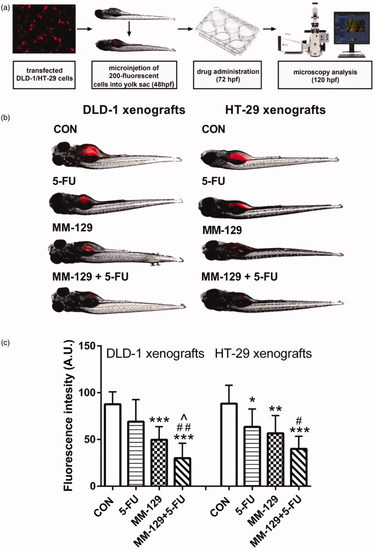
Schematic of xenograft assay and analysis of tumour development (a). Site-specific injection (yolk sac) of transfected (red) colon cancer cells (DLD-1 and HT-29) into 48 hpf zebrafish embryos and imaging analysis of tumour growth after 48 h of incubation with |