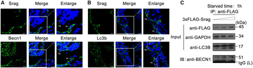
Srag coexpression with Becn1 and Lc3b and interaction between Srag and Becn1. (A, B) Coexpression analysis of Srag with Lc3b and Becn1. Immunofluorescence analysis of testis samples in serial sections using anti-Lc3b, anti-Srag, or anti-Becn1 antibody, respectively, then followed by FITC-conjugated ImmunoPure goat anti-rabbit IgG (green). The coexpression signals were detected in the cytoplasm of Sertoli cells (Sn) and spermatogonia (Sg) in testis. Lc3b-II puncta were detected in Sertoli cells and spermatogonia in testis (white arrows). The nuclei were stained by DAPI (blue). Images were captured using confocal microscopy. The enlarged images originated from the regions with white squares. Scale bar: 10 μm. (C) LC3B-II upregulation by srag overexpression and Co-IP between Srag and BECN1. HEK293T cells were transfected with an increasing amount of 3xFLAG-Srag (0, 0.5, and 1 μg). pcDNA3.0 was added for an equal amount DNA in each well. Western blot analysis indicated that LC3B-II is upregulated by Srag overexpression in a dose-dependent manner under starvation condition. Co-IP analysis indicated interaction between Srag and BECN1. Cell lysates were analyzed by western blotting with the anti-LC3B and anti-FLAG antibodies. GAPDH was used as an internal control.
|
