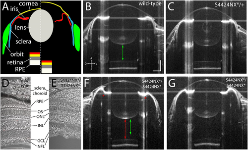
Figure 5. (A) Schematic showing wild-type (left of dashed line) and lrp2 mutant eye morphology (right of dashed line) as visualized by OCT. In lrp2 mutant eyes the retina is further away from the lens, and there is a larger gap between lens and cornea. Note the concavity of the iris in the mutant eye. (B) 2 mpf wild-type eye. Green arrow shows the distance from the back of the lens to the front of the retina, a proxy for vitreous chamber depth. Scale bar = 300 μm with the same scale used throughout the figure. (C) lrp2 S4424N*/+ heterozygous eye shows similar metrics to the wild-type eye. (D,E) Histological sections show normal lamination of wild-type and S4424NX* homozygous eyes (RPE, retinal pigment epithelium; OS, outer segments; ONL, outer nuclear layer; INL, inner nuclear layer; GCL, ganglion cell layer; NFL, nerve fiber layer). (F,G) lrp2 S4424N*/S4424N* homozygous eyes (left and right, respectively) show enlargement with glaucomatous phenotypes. The iris bulges inward (asterisk) and the vitreous chamber depth (red arrow) is greater than in the sibling wild-type from panel (B) (green arrow).
|
