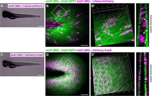
Distribution pattern of the mesenchymal cells in larval fins. (A) Image of the whole body and (B) the median fin fold of the transgenic (TG) larva expressing Lifeact-mCherry under the 5xand1(MC) promoter. The white dot line shows the outline of median fin fold. (B) Localization pattern of mesenchymal cells (expressing Lifeact-mCherry: magenta) and actinotrichia (labeled by And1-GFP: green) in the larval fin. (B′) Magnified image of the white box in (B). Transverse section image of the white dotted line is shown in the right panel. Actin-rich pseudopodia interact with actinotrichia (white arrowheads). (C) Image of the whole body and (D) the median fin fold of TG larva expressing mCherry-CaaX under the and1(BK) promoter. The white dotted line shows the outline of the median fin fold. (D) Localization pattern of basal keratinocytes (expressing mCherry-CaaX: magenta) and actinotrichia (labeled by And1-GFP: green) in the larval fin. (D′) The magnified image of white box in (D). Transverse section image at the white dotted line is shown in the right panel. All larvae were at the stage of 3 dpf. MC, mesenchymal cell specific promoter designated as 2PΔepi in the previous paper (Lalonde et al., 2016); BK, basal keratinocyte specific promoter designated as 1.4 k and1 pro in the previous paper (Kuroda et al., 2018). Scale bars: 500 μm in (A,C), 50 μm in (B,D).
|
