
Fig. 1
- ID
- ZDB-FIG-201117-6
- Publication
- Wen et al., 2020 - Primary and secondary motoneurons use different calcium channel types to control escape and swimming behaviors in zebrafish
- Other Figures
- All Figure Page
- Back to All Figure Page
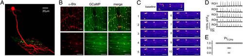
CaP 1°Mn occupies nearly all of the synapses formed on individual muscle cells. (A) Maximal projection of confocal images of a CaP (red) and an example target muscle with its associated receptor clusters (green). The CaP was labeled with tdTomato expression driven by the mnx1 promoter. Postsynaptic receptors were labeled with rapsyn-GFP driven by the α-actin promoter that sparsely labeled muscle cells. (B) Confocal Z-stack maximal projection (Top) and single focal plane (Bottom) images of approximately two segments showing colocalization of AChR labeling with α-Btx (red) and rapsyn-GCaMP6f basal fluorescence (green). (Scale bars, 20 μm.) (C) Example GCaMP6f fluorescence responses for five synapses in response to 0.2-Hz stimulation of the CaP (10 consecutive responses are shown). In this focal plane, ROIs 1 to 4 are located in the same muscle cell, while ROI 5 is on a different muscle. Scale bars, 15 μm. (D) Individual GCaMP6f responses for the five ROIs in response to 0.2-Hz stimulation of the CaP. (E) Overall distribution of Pr at 0.2 Hz measured for 59 ROIs from 16 fish. |