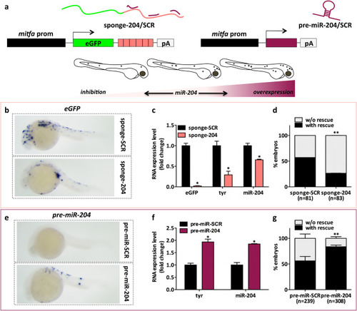
The constitutive modulation of miR-204 expression levels affects melanocyte content in embryos of the Tg(mitfa:BRAFV600E);p53−/−;mitfa−/− line. (A) (upper) Schematic representation of the miniCoopR vectors used for the inhibition and the overexpression of miR-204. On the left, miR-204 inhibition is achieved using a sponge construct that contains six imperfect binding sites for miR-204 downstream of eGFP coding sequence (miniCoopR-sponge-204). On the right, hsa-pre-miR-204 precursor is used to achieve the overexpression of mature miR-204 (miniCoopR-pre-miR-204). As negative controls, miniCoopR-sponge-SCR and miniCoopR-pre-miR-SCR are used, respectively. (lower) Cartoon summarizing the effects observed on embryos’ pigmentation. (B) eGFP mRNA detected by ISH 24 h after the injection of miniCoopR-sponge-SCR or miniCoopR-sponge-204 in 1-cell embryos. (C) eGFP, tyr and mature miR-204 levels measured by qRT-PCR at 7 dpf, upon the injection of miniCoopR-sponge-SCR or miniCoopR-sponge-204 in 1-cell embryos. (D) Percentage of 7dpf embryos showing rescued melanocytes, upon the injection of miniCoopR-sponge-SCR or miniCoopR-sponge-204 at the 1-cell stage. (E) pre-miR-204 RNA detected by ISH 24 h after the injection of miniCoopR-pre-miR-SCR or miniCoopR-pre-miR-204 in 1-cell embryos. (F) tyr and mature miR-204 levels measured by qRT-PCR at 7 dpf, upon the injection of miniCoopR-pre-miR-SCR or miniCoopR-pre-miR-204 in 1-cell embryos. (G) Percentage of 7 dpf embryos showing rescued melanocytes, upon the injection of miniCoopR-pre-miR-SCR or miniCoopR-pre-miR-204 at the 1-cell stage. Statistically significant differences are indicated with asterisks: *P<0.05, **P<0.01.
|
