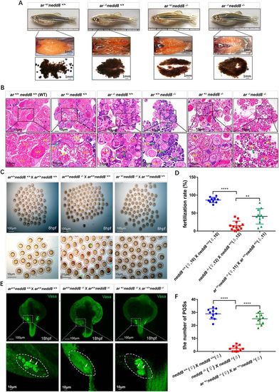
Loss of ar partially rescues nedd8−/− ovarian function. (A) Morphological comparison of ovaries and eggs from ar+/−nedd8+/+, ar−/−nedd8+/+, ar+/−nedd8−/− and ar−/−nedd8−/− female zebrafish at 4 mpf. Compared with the ovaries of the ar+/+nedd8−/− zebrafish (i.e. nedd8−/−; Fig. 1D), the ovaries of the ar+/−nedd8−/− zebrafish were filled with more oocytes in the midvitellogenic stage. (B) Histological analyses of the ovaries from female zebrafish with different genotypes at 4 mpf. Compared with oogenesis in the ar+/+nedd8−/− ovaries, oogenesis in ar+/−nedd8−/− ovaries was relatively normal. Green arrows indicate oocytes in different developmental stages. Bottom panels show magnification of the boxed areas in top panels. (C) Whole-mount images of embryos produced by different matings: ar+/+nedd8+/+ (♀)×ar+/+nedd8+/+ (♂); ar+/+nedd8−/− (♀)×ar+/+nedd8+/+ (♂); and ar+/−nedd8−/− (♀)×ar+/+nedd8+/+ (♂). The fertilization rate (number of fertilized eggs/total eggs laid×100) of the ar+/+nedd8−/− (♀)×ar+/+nedd8+/+ (♂) eggs was significantly lower than that of the ar+/−nedd8−/− (♀)×ar+/+nedd8+/+ (♂) eggs. Red arrows indicate degenerated embryos; green arrows indicate normal embryos. (D) Quantitation of the fertilization rates of different matings: ar+/+nedd8+/+ (♀)×ar+/+nedd8+/+ (♂) (n=10); ar+/+nedd8−/− (♀)×ar+/+nedd8+/+ (♂) (n=12); and ar+/+nedd8−/− (♀)×ar+/+nedd8+/+ (♂) (n=11). (E) Immunofluorescent staining of Vasa in PGCs of embryos (n=40) at 18 hpf from different matings: ar+/+nedd8+/+ (♀)×ar+/+nedd8+/+ (♂); ar+/+nedd8−/− (♀)×ar+/+nedd8−/− (♂); and ar+/−nedd8−/− (♀)×ar+/+nedd8−/− (♂). (F) The scatterplots represent PGC numbers in ar+/+nedd8+/+, ar+/+nedd8−/− and ar+/+nedd8−/−×ar+/− nedd8−/− embryos at 18 hpf. The numbers of PGCs were counted using a Leica dissection microscope based on the immunofluorescent density. EV, early vitellogenic stage; FG, full-grown stage; MV, midvitellogenic stage; PG, primary growth stage; PV, previtellogenic stage. Data are mean±s.e.m. **P<0.01, ****P<0.0001 (unpaired Student's t-test).
|
