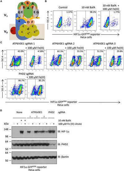
Iron supplementation restores HIF-1α levels to normal following ATP6V0E1 inhibition in HeLa cells. (A) Schematic diagram of the multimeric V-ATPase complex. (B) Chemical inhibition of V-ATPase by 10 nM BafA treatment for 24 h, increased HIF-1α levels in HIFα-GFPODD reporter cells. Treatment with 100 μM Fe (III) citrate significantly reduced the elevated HIF-1α levels associated with loss of ATP6V0E1 (1 × 10⁶ cells per sample harvested and analyzed; N = 2). (C) Knock-down of ATP6V0E1 subunit with three different CRISPR-Cas9 guides resulted in significant upregulation of HIF-1α levels in HIFα-GFPODD reporter cells. Co-treating cells with 100 μM Fe (III) citrate led to a reduction in HIF-1α levels across the three ATP6V0E1 depleted cells. phd2 was knocked down as a control and treatment with 100 μM Fe (III) citrate did not result in reduction of HIF-1α levels (1 × 10⁶ cells per sample harvested and analyzed; N = 2). FACs plot shown is a representative image of two biological repeats performed. (D) Immunoblot analysis for HIF-1α and PHD2 levels in HIFα-GFPODD reporter cells with either ATP6V0E1 or PHD2 depleted or treated with 10 nM BafA. The cells were treated with 100 μM Fe (III) citrate for 24 h. β actin was used as a control. Results validated findings observed by flow cytometry, whereby HIF-1α levels were upregulated following ATP6V0E1 knock-down or inhibition and levels were re-normalized upon Fe (III) citrate treatment. Treatment of Fe (III) citrate in PHD2 depleted cells did not alter HIF-1α levels. All experiments were performed in biological duplicate.
|
