
Fig. 2
- ID
- ZDB-FIG-200902-17
- Publication
- Peskin et al., 2020 - Notochordal Signals Establish Phylogenetic Identity of the Teleost Spine
- Other Figures
- All Figure Page
- Back to All Figure Page
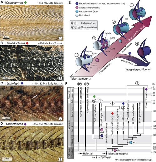
Patterning of the Spine and the Evolution of the Teleost Vertebral Centrum Reconstructing the changes in spine patterning and morphology during the evolution of teleosts. (A) The earliest members of stem teleosts lacked a vertebral centrum, and instead retained a functional notochord supporting the arches and spines. This is illustrated by the stem pachycormiform †Orthocormus roeperi; see also (E) state “0.” (B) †Pholidoctenus serianus illustrates a more derived condition present among some pholidophoriforms, with diplospondylous vertebral centra formed by hemi-chordacentra; see also (E) state “1.” (C) †Leptolepis coryphanoides illustrates a major transformation within the spine of stem teleosts, characterized by the appearance of a thin hourglass-shaped autocentrum surrounding a ring-like chordacentrum; see also (E) state “2.” (D) Subsequently, the autocentrum incorporated a thickened lateral wall, and a series of ornaments, such as crest, grooves and pits, as illustrated by the oldest fossil member of the teleost crown group †Anaethalion sp., a condition retained in, and defining all living teleosts; see also (E) state “3.” (E) Reconstructed scenario of the evolution of the teleosteomorph spine—from a basal functional notochord supporting the arches and spines—leading to a chordacentrum plus autocentrum in recent teleosts, or to a chordacentrum plus arcocentrum among extinct aspidorhynchiforms. (F) Consensus-phylogenetic hypothesis of the total-group teleosts or teleosteomorphs mapped on the geological timescale. The phylogenetic hypothesis was adapted from Arratia et al. (2019). As outgroup taxa, the analysis included five members of Holostei based on a total of 198 morphological characters. |