FIGURE

Figure 5

ID
ZDB-FIG-200826-12
Publication
Cotti et al., 2020 - More Bone with Less Minerals? The Effects of Dietary Phosphorus on the Post-Cranial Skeleton in Zebrafish
Other Figures
All Figure Page
Back to All Figure Page
Figure 5

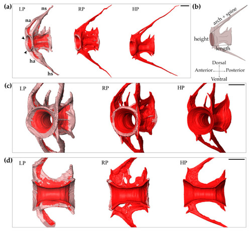
Increased bone formation in LP zebrafish after two months of dietary treatment. Synchrotron X-ray tomographic microscopy scans reveal an increased amount of non-mineralised matrix in the vertebral body and arches of low P diet (LP) treated animals compared to controls (RP) and high P diet (HP) treated individuals. (a) Lateral view of the 10th caudal vertebral body, neural (na) and haemal (ha) arches and their spines (ns, hs, respectively). Vertebral endplates are indicated by black arrowheads. Scale bar: 100 µm. (b) Schematic representation of the measured parameters: vertebral body length, vertebral body height, length of arch plus spine. (c) Frontal view of vertebrae. Scale bar: 100 µm. (d) Virtual sagittal sections of the vertebral bodies. Scale bar: 100 µm. Non-mineralised bone: pink; mineralised bone: red.

Expression Data

Expression Detail
Antibody Labeling
Phenotype Data

Phenotype Detail
Acknowledgments
This image is the copyrighted work of the attributed author or publisher, and ZFIN has permission only to display this image to its users. Additional permissions should be obtained from the applicable author or publisher of the image. Full text @ Int. J. Mol. Sci.