
Fig. 3
- ID
- ZDB-FIG-200728-56
- Publication
- Rago et al., 2019 - MicroRNAs Establish the Right-Handed Dominance of the Heart Laterality Pathway in Vertebrates
- Other Figures
- All Figure Page
- Back to All Figure Page
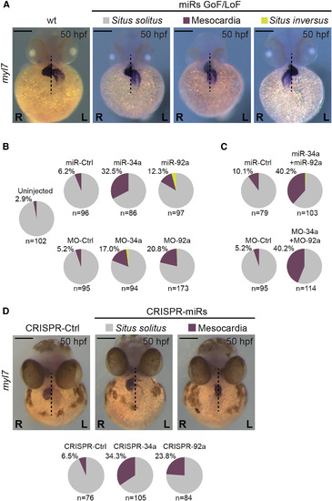
miRNAs Regulate Heart Positioning Zebrafish embryos were injected with synthesized miRNAs or MOs at 1–2 cell stage and collected at at 50 hpf to assess heart laterality. (A) After in situ hybridization for myl7 (cardiac myosin light chain 7) embryos were classified into three phenotypes: Situs solitus (normal condition, right heart looping after displacement of the posterior pole to the left), Mesocardia (heart in the middle), or Situs inversus (left heart looping after displacement of the posterior pole to the right). WT condition shows Situs solitus. (B) Pie charts represent the percentage of left/right displacement of the posterior pole from the midline or mesocardia found after miRNAs GoF (miR−) or downregulation (MO−). (C) Similar pie charts obtained after simultaneous GoF (upper) or knocking down (lower) of miR-34a and miR-92a. (D) Embryos injected at 1–2 cell stage with sgRNAs targeting the miRNA sequences analyzed at 50 hpf to assess heart laterality. Only normal position (Situs solitus) and mesocardia were observed. Percentage of mesocardia and number of embryos (n) is noted. (L), left; (R) right sides. Scale bar: 250 μm. See also Figure S3. |
Reprinted from Developmental Cell, 51, Rago, L., Castroviejo, N., Fazilaty, H., Garcia-Asencio, F., Ocaña, O.H., Galcerán, J., Nieto, M.A., MicroRNAs Establish the Right-Handed Dominance of the Heart Laterality Pathway in Vertebrates, 446-459.e5, Copyright (2019) with permission from Elsevier. Full text @ Dev. Cell