
FIGURE
Figure 2
- ID
- ZDB-FIG-200723-4
- Publication
- von der Heyde et al., 2020 - Translating GWAS-identified loci for cardiac rhythm and rate using an in vivo image- and CRISPR/Cas9-based approach
- Other Figures
- All Figure Page
- Back to All Figure Page
Figure 2
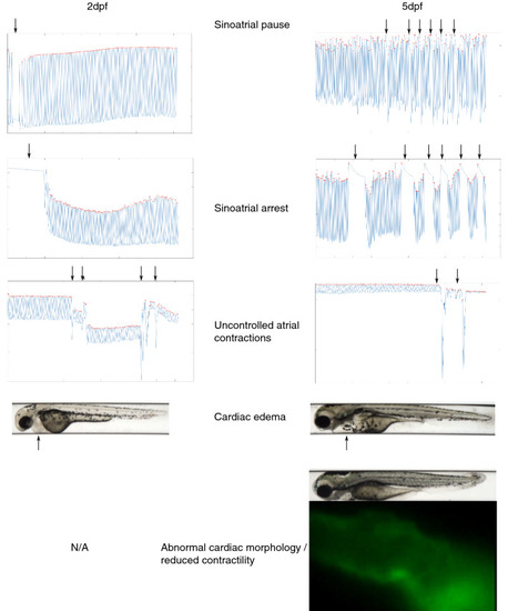
Visualization of cardiac rhythmic or other abnormalities, highlighted by arrows. Sinoatrial pauses were defined as the atrium ceasing to contract for > 3 × the median inter beat interval of the embryo. A sinoatrial arrest was defined as an event where the atrium stopped contracting for > 2 s. Abnormal cardiac morphology was defined as the atrium appearing as a tube-like structure, and impaired cardiac contractility was defined as the atrium vibrating rather than contracting. |
Expression Data
Expression Detail
Antibody Labeling
Phenotype Data
Phenotype Detail
Acknowledgments
This image is the copyrighted work of the attributed author or publisher, and
ZFIN has permission only to display this image to its users.
Additional permissions should be obtained from the applicable author or publisher of the image.
Full text @ Sci. Rep.