
FIGURE
Fig. S1
Fig. S1
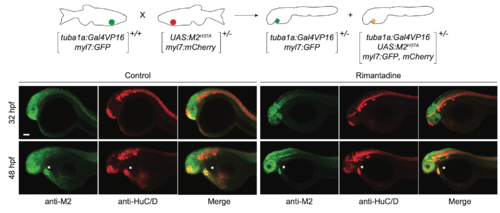
Neuron-specific expression of M2H37A. Representative maximum intensity projection micrographs of heterozygousTg(tuba1a:Gal4VP16;UAS:M2H37A;myl7:GFP, mCherry) embryos immunostained for the M2-derived channel (green) or HuC/D (red), an early neuronal marker. Asterisks denote residual fluorescence from reporter genes expressed in the heart. Embryo orientations: lateral view, anterior left. Scale bar: 100 μm. |
Expression Data
Expression Detail
Antibody Labeling
Phenotype Data
Phenotype Detail
Acknowledgments
This image is the copyrighted work of the attributed author or publisher, and
ZFIN has permission only to display this image to its users.
Additional permissions should be obtained from the applicable author or publisher of the image.
Full text @ Development