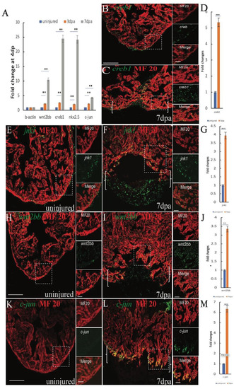
Induction of jnk1, creb1, wnt2bb and c-jun expression in injured cardiomyocytes during zebrafish heart regeneration (A) RT-qPCR results showing changes in wnt2bb creb1 nkx2.5 and c-jun expression at 4 and 7 dpa. (B,C) Confocal microscopy images showing creb1 expression during heart regeneration. At 7 dpa, creb1 expression expanded to cardiomyocytes adjacent to the injury site. Green, creb1; Red, MHC. (E,F) Confocal microscopy images of jnk expression after cardiac injury. The expression of jnk was low in uninjured ventricles, but at 7 dpa, jnk expression was enhanced and expanded into the new cardiomyocytes at the wound site (arrows). Green, jnk; Red, MHC. (H) Whereas wnt2bb expression was detectable on the uninjured ventricle (I), wnt2bb expression was enhanced in CMs adjacent to the injury site, and some non-CMs expressed wnt2bb at the apical edge of the wound at 7 dpa. Green, wnt2bb; Red, MHC. (K,L) Immunostaining analyses showed increased c-jun expression at the apical cell edges of the wounded heart and gradually increased at 7 dpa. Green, c-jun; Red, MHC. (D,G,J,M). Bar graph showing the fold changes in creb1, c-jun, jnk1, and wnt2bb expression in cardiomyocyte at 7 dpa compared to that observed in the uninjured heart. The data are presented as the means ± SEM, and significance was determined using Student’s t-test. n = 5. Brackets indicate the amputation area. Boxes correspond to the magnified region in the adjacent panels [Scale bars: 100 μm (left) and 25 μm (right)].
|
