FIGURE

Fig 6

ID
ZDB-FIG-200612-30
Publication
Weinschutz Mendes et al., 2020 - Expression of dlx genes in the normal and regenerating brain of adult zebrafish
Other Figures
All Figure Page
Back to All Figure Page
Fig 6

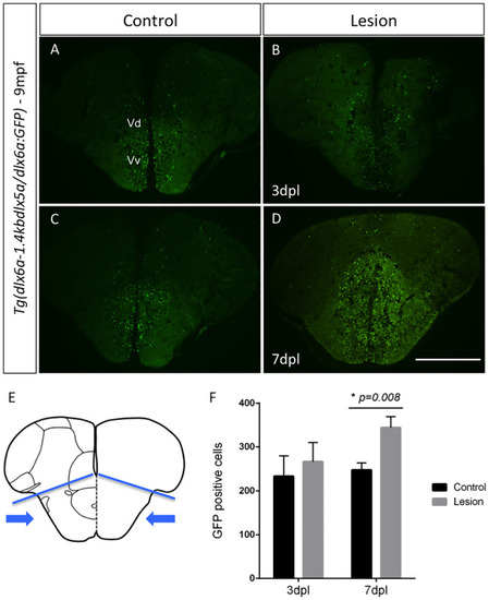
GFP labeling in Tg(dlx6a-1.4kbdlx5a/6a:GFP) adult zebrafish at 7 days post-lesion and cell quantification.

Expression of GFP in Tg(dlx6a-1.4kbdlx5a/6a:GFP) determined with a GFP antibody in controls (A and C) and regenerating brains at 3 dpl (B) and 7dpl (D). Schematic representation of the telencephalon where lesion is inflicted and areas used for cell counting of all GFP-positive cells (E) (area indicated by blue arrows bellow blue lines). Quantification of GFP+ cells in the regenerating brain at 3dpl and 7dpl (P = 0.008, N = 6) (F). Scale bar: 400 μm.

Expression Data
Gene:
Fish:
Condition:
Anatomical Term:
Stage: Adult

Expression Detail
Antibody Labeling
Phenotype Data
Fish:
Condition:
Observed In:
Stage: Adult

Phenotype Detail
Acknowledgments
This image is the copyrighted work of the attributed author or publisher, and ZFIN has permission only to display this image to its users. Additional permissions should be obtained from the applicable author or publisher of the image. Full text @ PLoS One