|
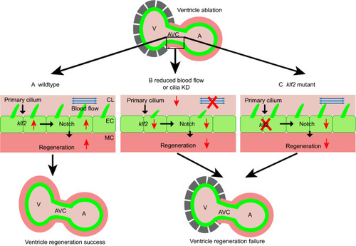
Diagrams of hemodynamic-responsive Klf2-dependant Notch activation in ventricle regeneration. (A) During normal ventricle regeneration process, primary cilia on the endocardial cells (green) sense the oscillatory blood flow (blue arrows), which leads to upregulation of the klf2 and subsequent activation of Notch signaling in the endocardium. This Notch activation is essential for myocardium (red) regeneration. (B) When blocking blood flow or impairing cilia development, endocardial klf2 expression and Notch signaling activation are inhibited, which lead to failure of ventricle regeneration. (C) In klf2 mutants, the lack of endocardial klf2 gene expression affects the activation of Notch signaling, and the damaged ventricle cannot regenerate. A, atrium; AVC, atrioventricular canal; CL, cardiac lumen; EC, endocardium; KD, knockdown; MC, myocardium; V, ventricle
|