|
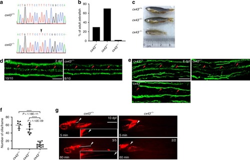
c<italic>x43</italic> null zebrafish have reduced motile cilia in ECs.a Electropherograms of the target sequences of the cx43 gDNA in WT and cx43−/− zebrafish. Arrow indicates the deletion of the T nucleotide. b Zebrafish (n = 71) at 2 months post-fertilization (mpf) from mating of cx43+/− zebrafish were genotyped for cx43. c Images of 3 mpf zebrafish with the indicated cx43 genotype. d WT or cx43−/− embryos at 2 dpf were immunostained with anti-acetylated-α-tubulin antibody, imaged with a confocal microscope and genotyped for cx43. Arrowheads represent motile cilia. Dorsal view anterior to the left. Scale bar = 20 μm. e Larvae at 8 dpf from mating of cx43+/− zebrafish were cut into cranial and caudal halves. The cranial half was used for cx43 genotyping, and the caudal half was coronally sectioned at a thickness of 14 μm and then processed for IF staining with anti-acetylated-α-tubulin antibody. Arrowheads represent motile cilia. Dorsal view anterior to the left. Scale bar = 30 μm. f Quantification of the number of cilia per frame in embryos in e. Mean ± SD. ****P < 0.0001 by one-way ANOVA with Tukey’s HSD post hoc test (cx43+/+ = 7 embryos; cx43+/− = 7 embryos; cx43−/− = 13 embryos; one frame per embryo). g Qdots were microinjected into the hindbrain ventricles of WT or cx43−/− zebrafish larvae at 10 dpf and then imaged at 5 and 60 min after the microinjection. Dashed lines indicate passive migration of Qdots. Arrowheads mark the caudal end of Qdot flow. Lateral view anterior to the left. Scale bar = 1 mm. Insets represent magnifications of the dotted areas.
|