
Fig. 2
- ID
- ZDB-FIG-200417-14
- Publication
- Sun et al., 2020 - Characterization of a nap1l1 transgenic reporter in zebrafish
- Other Figures
- All Figure Page
- Back to All Figure Page
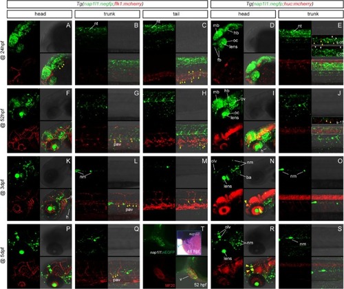
Early expression of nap1l1 transgenic line. Confocal imaging for the developmentally staged embryos or larvae from the out-cross of Tg(nap1l1:nEGFP)zs102 with the endothelial reporter line Tg(flk1:mCherry) and the neuronal reporter line Tg(huc:mCherry), respectively. (T) Confocal immunofluorescent imaging of the 52-hpf Tg(nap1l1:nEGFP)zs102 embryo with primary antibody MF20 (red). Inset demonstrates nap1l1-expressing cells within the developing heart (ventricle), which was analyzed by whole-mount in situ hybridization with antisense probe of nap1l1 mRNA. Abbreviations: ba, branchial arches; fb, forebrain; h, heart; hb, hindbrain; mb, midbrain; nm, neuromast; nt, neural tube; oc, optic cup; olv, olfactory vesicle; ov, otic vesicle; pav, paraxial vessel; v, ventricle. (For interpretation of the references to colour in this figure legend, the reader is referred to the web version of this article.) |
Reprinted from Gene, 735, Sun, S., Liu, Z., Li, X., Jia, J., Zhang, G., Yang, C., Jiang, Q., Zou, Y., Characterization of a nap1l1 transgenic reporter in zebrafish, 144388, Copyright (2020) with permission from Elsevier. Full text @ Gene