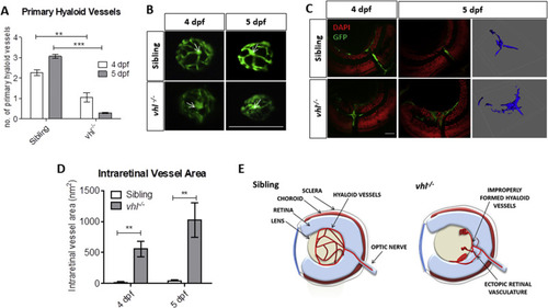
vhl−/− zebrafish larvae possess ocular vascular abnormalities at 4 and 5 dpf. A) Primary hyaloid vessel number at 4 and 5 dpf. vhl−/− larvae highlight a significant reduction in hyaloid branches at 4 and 5 dpf compared to sibling control. Data is representative of 3 biological replicates. N = 10 larvae per group per biological replicate. B) Fluorescent images of fli1:EGFP hyaloid vessels on sibling and vhl−/- dissected lenses at 4 and 5 dpf. White arrows illustrate central point from which primary branches are quantified. Scale bar = 200 μm. C) Confocal imaging revealed abnormal retinal neovascularisation (white arrows) at 4 and 5 dpf through the inner plexiform layer (IPL) and ganglion cell layer (GCL) compared to sibling control. Z-stacks confirmed aberrant vessel growth (blue) through the retina. Scale bar = 20 μm. D) Intraretinal vessel area is significantly increased in vhl−/− at both 4 and 5 dpf. N = 5 larvae per group E) Schematic of proposed ocular blood vessel formation in vhl−/− zebrafish larvae at 5 dpf.
|
