
Fig. 5
- ID
- ZDB-FIG-200327-3
- Publication
- Tiraboschi et al., 2020 - New insights into the early mechanisms of epileptogenesis in a zebrafish model of Dravet syndrome
- Other Figures
- All Figure Page
- Back to All Figure Page
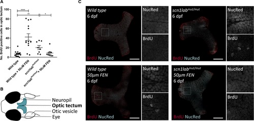
Proliferation of cells in the optic tectum of scn1labmut/mut larvae. A, At 6 days postfertilization (dpf), scn1labmut/mut larvae showed an increased number of proliferating cells in the optic tectum, as measured by BrdU‐positive cells in 30‐μm‐thick optical sections. Incubation of larvae in 50 μmol/L of fenfluramine (FEN) from 3 dpf onward resulted in an increased number of BrdU‐positive cells in wild‐type (WT) larvae at 6 dpf, while reducing the number to untreated WT levels in the scn1labmut/mut larvae. Statistical significance was calculated using multiple unpaired, two‐tailed t tests. * P < .05, ** P < .01, **** P < .0001. B, Cartoon representation of the head of a 6‐dpf larva, indicating the position of the optic tectum where proliferation was measured. C, Representative z‐slices of masked optic tecta with BrdU staining of WT and scn1labmut/mut larvae at 6 dpf, with or without prior incubation in 50 μmol/L of FEN from 3 dpf onward. Squares indicate the location of the zoomed‐in, split‐channel images to the right. Scale bars = 50 μm |
| Fish: | |
|---|---|
| Condition: | |
| Observed In: | |
| Stage: | Day 6 |