Fig. 4
- ID
- ZDB-FIG-200326-28
- Publication
- Yan et al., 2019 - Visualizing Engrafted Human Cancer and Therapy Responses in Immunodeficient Zebrafish
- Other Figures
- All Figure Page
- Back to All Figure Page
|
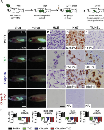
Combination Treatment of Temozolomide and Olaparib PARP Inhibitor Reduces Growth of Human RMS Cells in Engrafted prkdc−/−, il2rga−/− Zebrafish Schematic of experimental design (A). Merged fluorescence and brightfield images of prkdc−/−, il2rga−/− animals engrafted with EGFP+ RD cells (B). Whole-animal imaging of engrafted animal at 7 dpt (prior to drug administration, B, left) and 28 dpt (after three cycles of drug dosing, B, right). Hematoxylin and eosin (C), Ki67 (D), and TUNEL (E) stained sections of fish engrafted with RD RMS cells. The average percentage of positive cells ± standard deviation is noted (n = 5 fish/treatment). Quantification of relative RMS growth after drug administration in RD, SMS-CTR, Rh41, and Rh30 (F) cells. ∗p < 0.05, ∗∗p < 0.01, ∗∗∗p < 0.001, Student’s t test. Not significant (NS). Scale bar represents 0.25 cm (B) and 50 μm (C–E). Not applicable (NA). See also Figure S4. |
Reprinted from Cell, 177(7), Yan, C., Brunson, D.C., Tang, Q., Do, D., Iftimia, N.A., Moore, J.C., Hayes, M.N., Welker, A.M., Garcia, E.G., Dubash, T.D., Hong, X., Drapkin, B.J., Myers, D.T., Phat, S., Volorio, A., Marvin, D.L., Ligorio, M., Dershowitz, L., McCarthy, K.M., Karabacak, M.N., Fletcher, J.A., Sgroi, D.C., Iafrate, J.A., Maheswaran, S., Dyson, N.J., Haber, D.A., Rawls, J.F., Langenau, D.M., Visualizing Engrafted Human Cancer and Therapy Responses in Immunodeficient Zebrafish, 1903-1914.e14, Copyright (2019) with permission from Elsevier. Full text @ Cell

