
Fig. 1-S5
- ID
- ZDB-FIG-200325-15
- Publication
- Wee et al., 2019 - A bidirectional network for appetite control in larval zebrafish
- Other Figures
- All Figure Page
- Back to All Figure Page
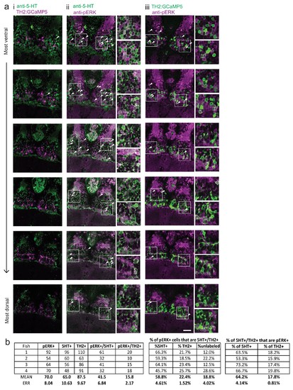
Food deprivation-induced activity in caudal hypothalamus monoaminergic neurons.( a) Dopaminergic neurons are labeled in Tg(TH2:GCaMP5) fish. These animals were food-deprived for 2 hr and then co-stained with anti-5-HT (to label serotonergic neurons) and anti-pERK antibodies in order to quantify food deprivation-induced activity in both cell types. Each row shows a different z-plane, moving from ventral to dorsal. (i) There is minimal overlap between Tg(TH2:GCaMP5)-positive cells (magenta) and 5-HT labeling (green). There is higher overlap of anti-pERK staining (magenta) with (iii) 5-HT-positive cells (green) as compared to (ii) Tg(TH2:GCaMP5)-positive cells (green). White arrows point to examples of overlapping cells. White boxes indicate region shown in insets. Scale bar = 20 μm. Full z-stacks for (ii) pERK overlap with anti-5-HT staining ( Video 2) and (iii) TH2:GCaMP5 expression ( Video 3) are also provided. ( b) Quantification of overlap between pERK-positive cells and anti-5-HT staining or Tg(TH2:GCaMP5) expression. Other cH cell types, including histaminergic neurons ( Chen et al., 2016) are not labeled. Fish one corresponds to the fish shown in ( a). |