
Figure 1
- ID
- ZDB-FIG-200306-95
- Publication
- Coolen et al., 2020 - Mosaic Heterochrony in Neural Progenitors Sustains Accelerated Brain Growth and Neurogenesis in the Juvenile Killifish N. furzeri
- Other Figures
- All Figure Page
- Back to All Figure Page
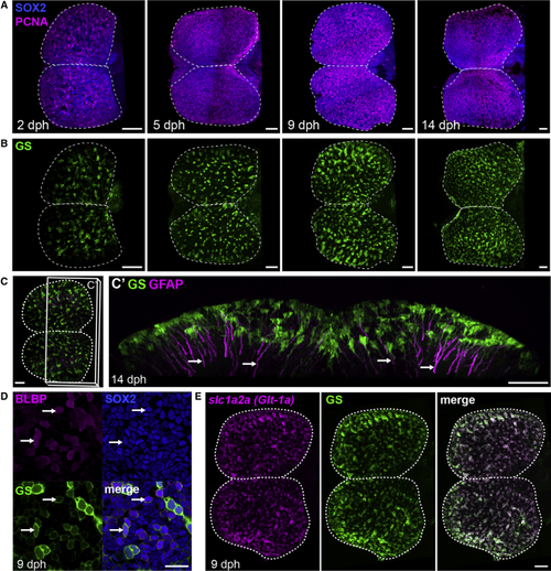
Apical Progenitors at the Pallial Surface of the Killifish Larval Pallium (A) Dorsal 3D views of killifish pallium (anterior left) at 2, 5, 9, and 14 days post-hatching (dph) with a whole-mount immunostaining for Sox2 (blue) and PCNA (magenta) highlighting neural progenitors. A dotted line contours the two pallial hemispheres. (B) Dorsal 3D views of the same brains as in (A), showing immunostaining signal for GS (green) to identify RGs. (C) Immunostaining for GS (green) and GFAP (magenta) at 14 dph indicating GFAP-enriched processes (arrows). Shown in (C) is a dorsal 3D view, and in (C’) is a frontal view of the 3D reconstruction with a transverse hemisection along the plane shown in (C). (D) Triple immunostaining for BLBP, Sox2, and GS at 9 dph. Images show high magnifications of the pallial surface on a single optical z-plane. Arrows point to RG cells (GS+BLBP+Sox2+). Scale bar, 20 μm. (E) ISH for See also |