FIGURE

Figure 3

ID
ZDB-FIG-200306-17
Publication
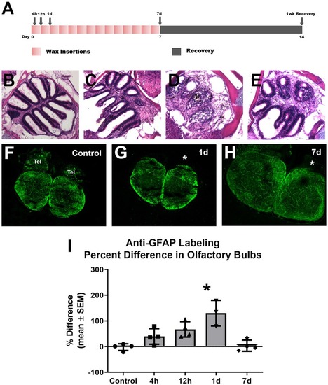
Scheib et al., 2020 - Zebrafish Astroglial Morphology in the Olfactory Bulb Is Altered With Repetitive Peripheral Damage
Other Figures
All Figure Page
Back to All Figure Page
Figure 3

Gross analysis with Z-stack images of anti-GFAP labeling in the olfactory bulb following repetitive peripheral damage. (A) Illustration of the treatment paradigm shows that fish receiving repeated wax plug insertions were analyzed at various survival times during wax treatment and after a recovery period. Images of olfactory organs show typical rosette morphology in control fish (B), significant damage at 1 day (C) and 7 days (D) of wax plug insertions, and return to control morphology with 1 week of recovery (E) after removal of the plug. (F) Untreated control fish had typical anti-GFAP labeling in all layers of both olfactory bulbs. Tel = Telencephalon, n = 4. (G) After 1 day of repetitive peripheral damage to the olfactory organ, the affected bulb (asterisk) appeared to have slightly more anti-GFAP labeling than the internal control bulb, n = 3. (H) By 7 days, there was no noticeable difference in labeling between bulbs, and anti-GFAP labeling in the affected bulb (asterisk) appeared at control levels, n = 4. Scale bar = 100 μm for all.(I) A comparison of anti-GFAP labeling between bulbs showed a significant difference at 1 day of repetitive peripheral damage when compared to controls but no difference at 7 days of damage. *p < 0.05.

Expression Data

Expression Detail
Antibody Labeling
Phenotype Data

Phenotype Detail
Acknowledgments
This image is the copyrighted work of the attributed author or publisher, and ZFIN has permission only to display this image to its users. Additional permissions should be obtained from the applicable author or publisher of the image. Full text @ Front. Neuroanat.