FIGURE

Figure 1.

ID
ZDB-FIG-200302-21
Publication
Son et al., 2020 - Dopaminergic Co-Regulation of Locomotor Development and Motor Neuron Synaptogenesis is Uncoupled by Hypoxia in Zebrafish
Other Figures
All Figure Page
Back to All Figure Page
Figure 1.

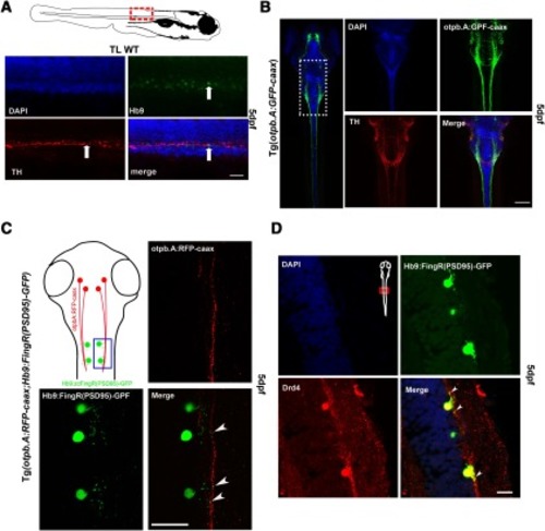
Dopaminergic synapses to spinal cord motor neurons. A, Confocal images, lateral views of zebrafish spinal cord at 5 dpf, dorsal to top; scale bar: 10 µm. DAPI staining of cell nuclei; anti-Hb9 immunohistochemistry in motor neurons; anti-TH immunohistochemistry of DDT; and double-labeling anti-Hb9 anti-TH shows co-localization (arrows). B, Confocal images, dorsal views of zebrafish at 5 dpf, rostral top. Left panel shows entire larva with DDT labeled by GFP in Tg(otpb.A:EGFP-caax) line. Higher magnification (scale bar: 50 µm) shows DAPI staining of cell nuclei; GFP labeling of DDT; anti-TH labeling of TH; and co-localization of TH and GFP in DDT. C, Tg(otpb.A:TRFP-caax); Tg(Hb9:FingR(PSD95)-GFP) larvae, confocal images, dorsal views, of spinal cord at 5 dpf (scale bar: 10 µm). FingR localization of PSD95, expressed in motor neurons, of synapses can be seen neighboring DDT labeled by RFP (arrowheads). D, Anti-D4R labeling co-localizes with FingR GFP expression (scale bar: 10 µm).

Expression Data

Expression Detail
Antibody Labeling
Phenotype Data

Phenotype Detail
Acknowledgments
This image is the copyrighted work of the attributed author or publisher, and ZFIN has permission only to display this image to its users. Additional permissions should be obtained from the applicable author or publisher of the image. Full text @ eNeuro