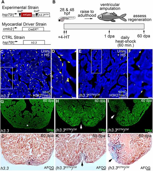
Myocardial H3K27 tri-methylation is required for zebrafish heart regeneration in vivo. (A) Schematics depicting driver and inducible transgenes used for Cre/loxP-mediated myocardial-specific expression of Tg(hsp70l:h3.3) (wild-type control) or Tg(hsp70l:h3.3K27M)CM (mutant) during the regenerative window. (B) Schematic depicting the experimental strategy for heart regeneration analyses. Double-transgenic embryos were exposed to 4-HT at 24 h post-fertilization (hpf) and 48 hpf to recombine the mutant transgene specifically in the myocardium. Zebrafish were raised to adulthood and their ventricles resected followed by a 24 h recovery period and daily 1 h heat-shocking for 60 days prior to analysis. (C-F) Sections of uninjured zebrafish ventricles co-immunostained for the cardiomyocyte marker, myosin heavy chain (blue) and H3K27me3 (yellow). Boxed regions in C and E are shown in D and F. Myocardial and non-myocardial cells displayed strong H3K27me3 signals in Tg(hsp70l:h3.3) hearts (C,D; n=5/5 hearts). By contrast, myocardial H3K27me3 signals failed to be detected in Tg(hsp70l:h3.3K27M)CM hearts, whereas non-myocardial signals were preserved (E,F; n=5/5 hearts). (G-L) Representative cardiac sections from 60 dpa heat-shocked Tg(hsp70l:h3.3) control (G,J) and Tg(hsp70l:h3.3K27M)CM (H,I,K,L) animals evaluated by immunofluorescence for the myocardial marker TPM (green; G-I) or AFOG staining (J-L). Whereas control animals robustly regenerated myocardium (asterisks in G,J; n=9/9 hearts), Tg(hsp70l:h3.3K27M)CM hearts failed to regenerate new muscle (arrows in H and I; n=9/11) with either ventricular wall deficits (arrow in K; n=4/11) or apparent collagen deposits indicative of scarring (arrow in L; n=5/11). Scale bars: 50 µm in C,E; 10 µm in D,F; 100 µm in G-L.
