
Fig 7
- ID
- ZDB-FIG-200220-29
- Publication
- Messerli et al., 2020 - Adaptation mechanism of the adult zebrafish respiratory organ to endurance training
- Other Figures
- All Figure Page
- Back to All Figure Page
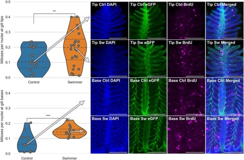
Top half: Data and images from the tips of the gills, bottom half: Data and images from the gill bases. Column 1: Plots of the number of mitoses per total number of nuclei in immunostained gill tips or bases (n = 6 for each group). After 3 weeks of training, the trained fish show a significantly higher number of dividing cells in their gills, compared to controls, both at the tips of the gills and at the base of them (tips: p = 0.0074, base p = 0.00084). The arrows point from the median value to the corresponding row of microscopy images. Columns 2-4: Staining of the nuclei in blue (DAPI), endothelium in green (eGFP) and mitoses in magenta (BrdU). Column 5: Composite image of the three channels. The microscopy images shown correspond to the median value of the data in the first column. Scale bar: 0.1 mm. **: p<0.01, lines within the plots show the quartiles of the respective distributions. |