FIGURE

Figure 2

ID
ZDB-FIG-200125-13
Publication
Chen et al., 2020 - Microarray Expression Profiling and Raman Spectroscopy Reveal Anti-Fatty Liver Action of Berberine in a Diet-Induced Larval Zebrafish Model
Other Figures
All Figure Page
Back to All Figure Page
Figure 2

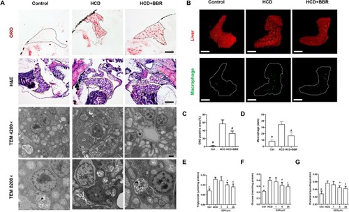
Effects of berberine (BBR) on hepatic steatosis in high-cholesterol diet (HCD)-induced zebrafish larvae. (A) The zebrafish larvae were treated with the control diet, HCD diet, and HCD plus BBR (5 μM) treatment. Hepatic steatosis was observed by whole-body Oil Red O (ORO) staining (line 1, refer to the black dotted, bar = 100 μm), H&E staining (line 2, refer to the yellow dotted, bar = 100 μm), and TEM (line 3, bar = 2μm; line 4, bar = 1 μm). m mitochondrion, n cell nucleus, LD lipid droplet. (B) L-plastin-labeled macrophages in Tg (fabp10a: dsRed) zebrafish. The statistical results of the positive area of ORO staining were shown in (C) and the quantity of macrophage in the liver of larval zebrafish in (D). Effects of BBR on the levels of triglyceride (E), glucose (F), and cholesterol (G) in livers of zebrafish larvae fed with HCD. #Compared with the HCD group, P < 0.05.

Expression Data

Expression Detail
Antibody Labeling
Phenotype Data
Fish:
Conditions:
Observed In:
Stage: Days 14-20

Phenotype Detail
Acknowledgments
This image is the copyrighted work of the attributed author or publisher, and ZFIN has permission only to display this image to its users. Additional permissions should be obtained from the applicable author or publisher of the image. Full text @ Front Pharmacol