FIGURE
Fig 7
- ID
- ZDB-FIG-191230-1358
- Publication
- Pazhakh et al., 2019 - β-glucan-dependent shuttling of conidia from neutrophils to macrophages occurs during fungal infection establishment
- Other Figures
- All Figure Page
- Back to All Figure Page
Fig 7
|
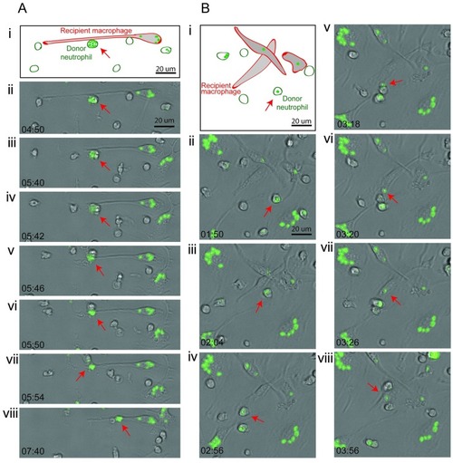
(A,B) Two sequences demonstrating neutrophil-to-macrophage shuttling of Alexa-Fluor-488–labeled zymosan particle between murine phagocytes in vitro. Panel (i) is a schematic showing the elongated, adherent recipient macrophage. Panels (ii–viii) are bright-field photomicrographs with green fluorescence channel overlaid, with time points indicated in min:s. Red arrow indicates the shuttled particle in donor neutrophil (panels ii–vi) and then, following shuttling, within the recipient macrophage (panels vii–viii). Stills from |
Expression Data
Expression Detail
Antibody Labeling
Phenotype Data
Phenotype Detail
Acknowledgments
This image is the copyrighted work of the attributed author or publisher, and
ZFIN has permission only to display this image to its users.
Additional permissions should be obtained from the applicable author or publisher of the image.
Full text @ PLoS Biol.

